


2014 kavli prize in Nanoscience
2014 Kavli
Prize in
Nanoscience
The Norwegian Academy of Science and Letters has decided to award the 2014 Kavli Prize in Nanoscience to
Thomas W. Ebbesen, Stefan W. Hell and Sir John B. Pendry.


“For their transformative contributions to the field of nano-optics that have broken long-held beliefs about the limitations of the resolution limits of optical microscopy and imaging.”
Committee Members
- Arne Brataas (Chair), The Norwegian University of Science and Technology, Norway
- Mildred Dresselhaus, Massachusetts Institute of Technology, USA
- Evelyn Hu, Harvard University, USA
- Klaus Kern, Max Planck Institute for Solid State Research, Germany
- Xie Yi, University of Science and Technology of China, China
Citation from the Committee
"Seeing" has often been the precursor to our cultural and scientific understanding of the world. Scientists have realized this from the time of Hooke and van Leeuwenhoek in the 17th century, when their innovations in optics opened up a new world at the micron scale.
"Seeing at the nanoscale" was long-considered to be limited in visible resolution by the finite wavelength of "light," so that only objects larger than ~ 200 nanometres could be imaged. This is about 100 times smaller than the diameter of a human hair. Other technologies have been developed to allow us to overcome these resolution limitations: for example, beams of electrons and scanning probe instruments. However, for ease of use and compatibility with biological specimens, optical microscopy has unparalleled advantages. Each of this year’s prize winners, through their different insights and routes, has independently advanced our ability to "see" nanostructures using "ordinary" light. This ability to see and image nanoscale objects is a critical prerequisite to further advances in the broader field of nanoscience. Thus, this year’s prize winners have not only advanced our understanding of nano-optics, but in addition, the application of these new insights into the imaging process in turn promises to have an enduring benefit to a wide range of fields ranging from physics and chemistry to the biological and biomedical sciences.
Thomas W. Ebbesen is recognized with the Kavli Prize in Nanoscience
“for the discovery of the extraordinary transmission of light through sub-wavelength apertures.”
"Common wisdom" tells us that objects cannot pass through openings that are much smaller than themselves. In fact, since the 1940s, the definitive reference for the behaviour of light transmitted through small holes in a metal sheet was Bethe’s work, which predicted that the light intensity would fall off dramatically as the radius of the hole diminished substantially below the wavelength of light. This limitation poses a real challenge to optics and imaging at very small dimensions. Ebbesen has shown that, on the contrary, there can be an extraordinary transmission of light through nano-fabricated holes in thin metal films. The sizes of those holes are far smaller than the wavelength of the light itself.
His experiments in 1998 yielded results that thus challenged all prior accepted theories of light propagation through small holes. The underlying reasons have to do with efficient re-radiation made possible through plasmons – a cooperative oscillation of electrons, particularly intense in nanoscale structures. Ebbesen’s understanding of the basic mechanism, and his implementation of different structures to enhance the focus, direction, and general control of the plasmonic enhancement have led to new means of increasing the efficiency, spatial focus of photonic devices, and sensitivity of optical sensors.
Stefan W. Hell is recognized with the Kavli Prize in Nanoscience
“for ground-breaking developments that have led to fluorescence microscopy with nanometre scale resolution, opening up nanoscale imaging to biological applications.”
Ernst Abbe demonstrated in 1873 that optical microscopy should not be able to discern features that are closer than half the wave-length of light. The Abbe limit became a cornerstone of optics that was not questioned for the next 120 years.
Hell met this challenge and overcame the diffraction limit by more than an order of magnitude. He accomplished this through understanding both the imaging mechanisms and the nature of what is being imaged. A key issue in the clarity of an image has always been distinguishing signal from a broad background of noise. By deeply understanding the composition of what is being imaged, be it biological or non-biological in nature, Hell showed how to control the background noise by strategically "shutting off" molecular transitions at the appropriate time. He calls this "shutting off" Stimulated Emission Depletion (STED): a technique that has now become accessible through instruments which he has helped to make commercially-available. Not only has STED-enabled imaging at dimensions far smaller than optical wavelengths for a broad class of materials, it has in particular made this a viable option for the life sciences. Remarkably, Hell’s techniques have made possible direct observation of dynamical processes in living cells at nanoscale resolution.
Sir John B. Pendry is recognized with the Kavli Prize in Nanoscience
“for developing the theory underlying new optical nanoscale materials with unprecedented properties, such as the negative index of refraction, allowing for the formation of ‘perfect lenses'.”
For many of us, our association with "optics" is with the corrective lenses we wear, which often have imperfections: the images that are not quite in focus or have "aberrations" or rainbow-like haloes associated with the images. The issues are associated with the ways in which light "bends" when going from air through lens material and into air again, and this is related to the index of refraction of those materials. Pendry has created a model for constructing a perfect lens, based on materials not normally found in nature. Such special materials have a negative index of refraction, as previously discussed by Veselago. It was Pendry’s insight to reinvestigate these ideas in the context of real materials such as silver, gold, and copper, and to formulate the guidelines for the eventual realization of perfect lenses. With a growing body of experimental validation of his work, he has further stimulated a plethora of activities extending these concepts all the way across the visible spectrum and beyond.
Pendry has helped to formulate rules on how to incorporate different kinds of materials (metals and dielectrics) with nanoscale structures to form larger scale "metamaterials" with exciting new optical properties that nature has not before provided us with. Thus, he encourages us to challenge our previously held ideas about the kinds of optical materials that we can engineer, promising dramatically improved levels of efficiency in light emission, storage, and sensing.
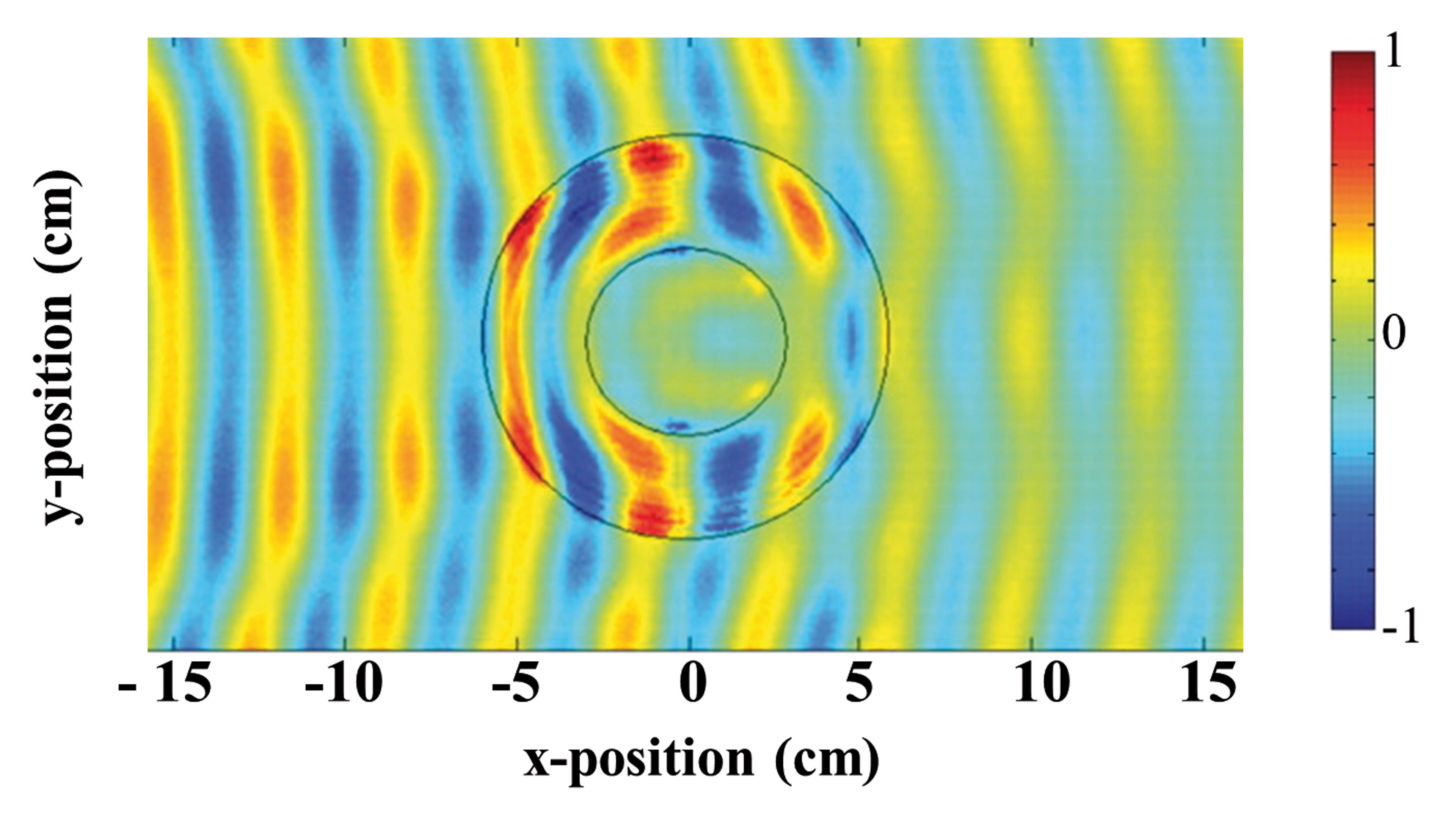
Electric field patterns of microwave radiation with a frequency of 8.5 GHz as it propagates through a metamaterial cloak (the region between the two black circles) wrapped around a copper cylinder (inner black circle). (Photo credit: Science 314, 977-980 [2006])
The 2014 Nanoscience Kavli Prize explained
The ability to image objects with visible light is at the heart of our perception of the world, and is essential in fields ranging from cancer diagnostics to astronomy. Up until only a few years ago, the resolution of optical imaging was thought to be limited by the wavelength of light, making it impossible to image nano-objects.
Elisa De Ranieri and Fabio Pulizzi
In making their award, the Kavli Prize Committee in Nanoscience has selected three scientists who have demonstrated how to break this resolution limit and how to use light to interact with matter with nanoscale precision, allowing the development of new and more efficient imaging techniques and photonic devices.
Thomas W. Ebbesen was primarily working on carbon nanostructures when he made an extraordinary observation in a different field. He noticed that light could be transmitted through a metal plate with a pattern of holes that were much smaller than the wavelength. The phenomenon was unexpected because it was assumed that, when a beam of light hits a hole smaller than its wavelength, it is affected by very poor transmission and strong diffraction.
More systematic experiments showed that this extraordinary optical transmission, as it was named in the seminal paper that reported the results in 1998, was only occurring for specific wavelengths, and there was a well-defined relationship between these wavelengths and the periodicity of the holes. The phenomenon was related to the interaction of the light beam with surface plasmons. These are collective electron states in a metal that can harvest the incident light at a wavelength determined by the geometry of the metallic surface, that is, the periodicity and shape of the holes. The effect of the plasmons was so strong that they could channel part of the light hitting the metal surface, and send it through the holes, resulting in a transmission higher than 100%.

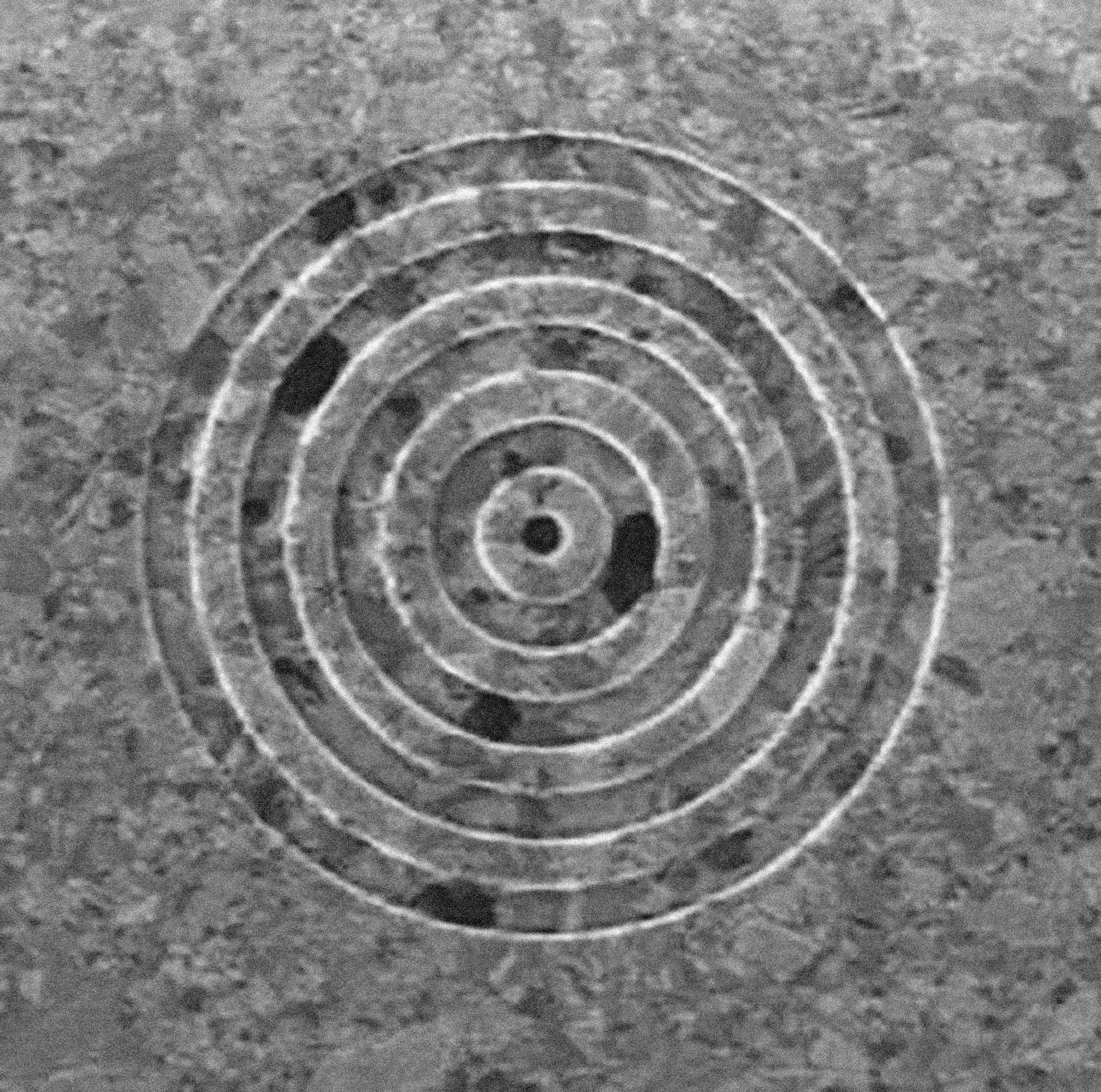
Focused ion beam micrograph image of the bull’s eye structure on a 300 nm-thick silver film used to demonstrated transmission through a subwavelength hole. (Photo credit: Science 297, 820-822 [2002])
Ebbesen kept working on this topic and shortly after he and his colleagues showed that light could also be transmitted through a single hole in a metal plate measuring only a couple of hundred nanometers in diameter. This worked as long as the hole was surrounded by a periodic structure of grooves in the metal that allowed a light beam to be coupled with the plasmons. What is more, not only can light be transmitted through such a small hole; it can also be beamed in a collimated ray. Ebbesen and co-workers studied a so-called bull’s eye structure, consisting in a periodic pattern of concentric grooves in a silver plate around a 200 nm hole. If the same structure is fabricated also on the output side of the hole, the light exits as in an almost parallel beam.
The extraordinary light transmission through sub-wavelength holes has several applications in diverse fields of photonics. Because of the dependence of the transmitted wavelength on the geometry of the metal plate, it is possible to design very selective optical filters or polarizers. The creation of collimated and very small beams can be used in photonic devices like lasers. Furthermore, it was shown that the fluorescence of a single molecule collected through one such hole is substantially enhanced. Finally, it is, in principle, possible to use the phenomenon to obtain very small features in lithography, the technique of forming patterns onto materials that allows subsequent formation of devices and circuits.
In the late 1980s, while working on his postgraduate project in Heidelberg, Stefan W. Hell became interested in ways to improve the resolution of confocal microscopy. In particular, he convinced himself that it would be possible to break the barrier of 200 nm resolution in optical microscopy imposed by Ernst Abbe’s law in 1873, assumed as a fundamental limit by the whole scientific community.
According to Abbe’s law, the optical resolution of a microscope is limited by the smallest spot at which a microscope objective can focus a laser beam. For example, in fluorescence microscopy, molecular dyes known as fluorophores are injected into a tissue and illuminated to produce an image. Fluorophores within the laser spot are all illuminated at the same time, resulting in a resolution which is at best 200 nm.
Hell realized that the best way to overcome the limitations set by Abbe’s law was to affect the electronic states of the fluorophores. In standard fluorescence microscopy, a laser beam is used to excite the fluorophores from their ground states to a state with higher energy. After a certain time, the fluorophores decay back to their ground state by the emission of light. In 1994, while working in Turku, Finland, Hell published a theoretical paper explaining the concept of what is now known as stimulated emission depletion (STED) microscopy. Beside the laser beam used in fluorescence microscopy, a second, donut-shaped beam was superimposed to the first one to force all fluorophores located outside the central hole of the donut back to the ground state without emitting light. By varying the intensity of the laser and the shape of the donut, it would be possible to improve the resolution, in principle to a few nanometers.

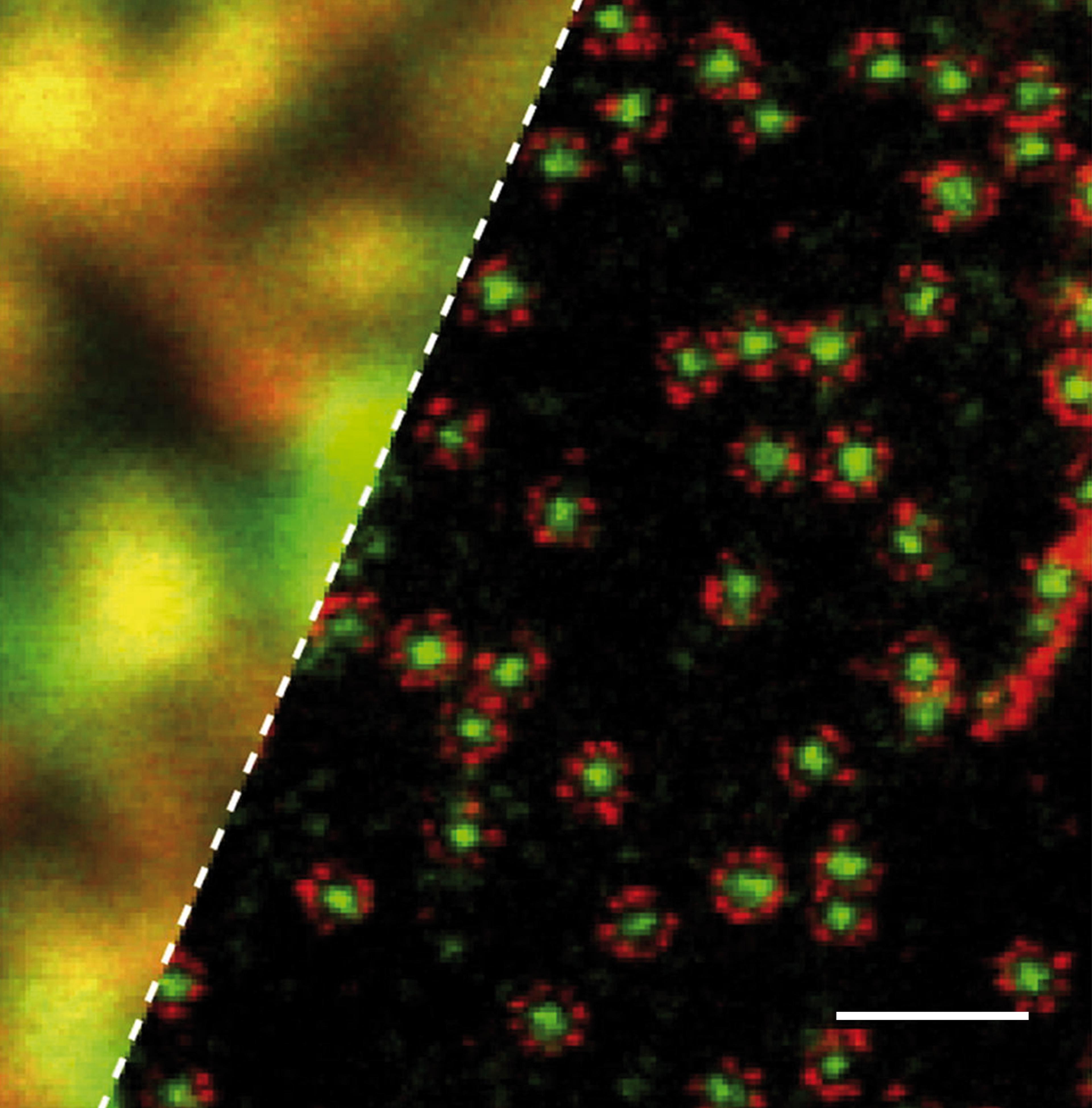
STED microscopy image of protein complexes (right), revealing a much higher resolution than conventional confocal microscopy (left). The scale bar is 500 nm. (Photo credit: Biophysical Journal 105, L01–L03 [2013])
The concept was intriguing but it had to wait a few years to be widely accepted. In 2000, Hell and co-workers published another paper, this time experimental, demonstrating the use of STED microscopy to obtain images with a resolution of just over 100 nm. In the following years, Hell’s research helped improving the resolution down to a few tens of nanometers and to develop other forms of microscopy, based on the concepts of STED, but necessitating much lower laser power. One such example is reversible saturable optical fluorescence transition (RESOLFT) microscopy.
Hell’s work has always been driven by the principle that the power of an optical microscope is ultimately determined by its resolution, and that the ability to distinguish details only a few nanometers apart would lead to essential information on how proteins, lipids, and even neurons interact with each other. A beautiful example of the success of his approach is the imaging, in 2012, of the brain of a living mouse with a 70 nm resolution.
John B. Pendry of Imperial College London designed artificial materials with properties not found in nature, and showed that such materials can be used to fabricate optical lenses with a resolution smaller than the wavelength of light, thus allowing imaging of objects with nanoscale dimensions.
The materials inspired by Pendry’s vision since the 1990s are called "metamaterials." They are composed by nanoscale metallic or dielectric elements, with a geometry that determines the optical properties. What is more, this geometry can be engineered to obtain the desired response to electric and magnetic fields. Pendry’s work also predicts metamaterials with a negative refractive index, a feature not found in nature, and resulting in dramatically different ways in which light “bends” as it traverses two different materials. Negative index materials were first experimentally demonstrated by David Smith in 2000.

Electric field patterns of microwave radiation with a frequency of 8.5 GHz as it propagates through a metamaterial cloak (the region between the two black circles) wrapped around a copper cylinder (inner black circle). (Photo credit: Science 314, 977-980 [2006])
In 1968, Victor Veselago predicted that a slab of material with negative refractive index can focus light. Building on this work, Pendry theoretically demonstrated in 2000 that such a slab not only can focus light, but also that it does so with sub-wavelength resolution, which cannot be achieved with standard lenses. Pendry named this slab of metamaterial a "perfect lens," because it can focus light of a given wavelength with unlimited resolution, if the index of refraction is exactly -1. This is due to the action of surface plasmons on both sides of the slab, as they amplify the components of the electromagnetic radiation - the so-called "near field" - that carries sub-wavelength information of the object. This is information that is normally lost in conventional optical lenses.
Lenses with sub-wavelength resolution built from silver metamaterials have been experimentally demonstrated in 2005 by two independent groups in Berkeley and Canterbury. Such devices can be applied in a vast range of technologies, from imaging of small biological structures such as DNA molecules, to manufacturing of electronic devices with smaller dimensions or optical data storage with increased information density.
The applications of metamaterials go beyond perfect lenses; they can also be used selectively to absorb different wavelengths of radiation, or to realize the "invisibility cloak," proposed by Pendry in the early 2000s. When shaped around an object, the metamaterial cloak can hide it from electromagnetic radiation, making it invisible to an observer. Cloacking is analogous to creating a mirage; light is bent in a way that our brain reconstructs. In 2006, Pendry, David Smith and co-workers demonstrated cloaking at microwave frequencies. Although cloaking has only been demonstrated for small objects and may not be the most promising application of metamaterials, the concept is interesting as it challenges our intuition and understanding of the behavior of light.
Professor Arne Brataas, of the Norwegian University of Science and Technology, and chairman of the Kavli Nanoscience Prize Committee, said: “Thomas W. Ebbesen, Stefan W. Hell, and John B. Pendry have independently advanced our ability to 'see’ nano-scale objects using visible light. They have greatly advanced our understanding of nano-optics and the applications of their insights promise to have an enduring benefit to a wide range of fields from physics and chemistry to the biological and biomedical sciences”.