


From birds to brains: My twisted (and very lucky) path to the fusiform face area
As told by Nancy Kanwisher
Mine is not one of those inspiring stories of people who found their way to science against all odds. I grew up in Woods Hole, Massachusetts, where science was handed to me on a platter, from the Children's School of Science to the summer courses one could just walk into uninvited, to the library of the Marine Biology Labs which was open at all hours every day of the year, to the Friday Evening Lectures, the place to see and be seen in town.
My first publication, on the physiology of diving birds, was co-authored with my dad a field biologist at the Woods Hole Oceanographic Institution, and his then-student Geir Wing Gabrielsen, now of the Norwegian Polar Institute (Figure 1).

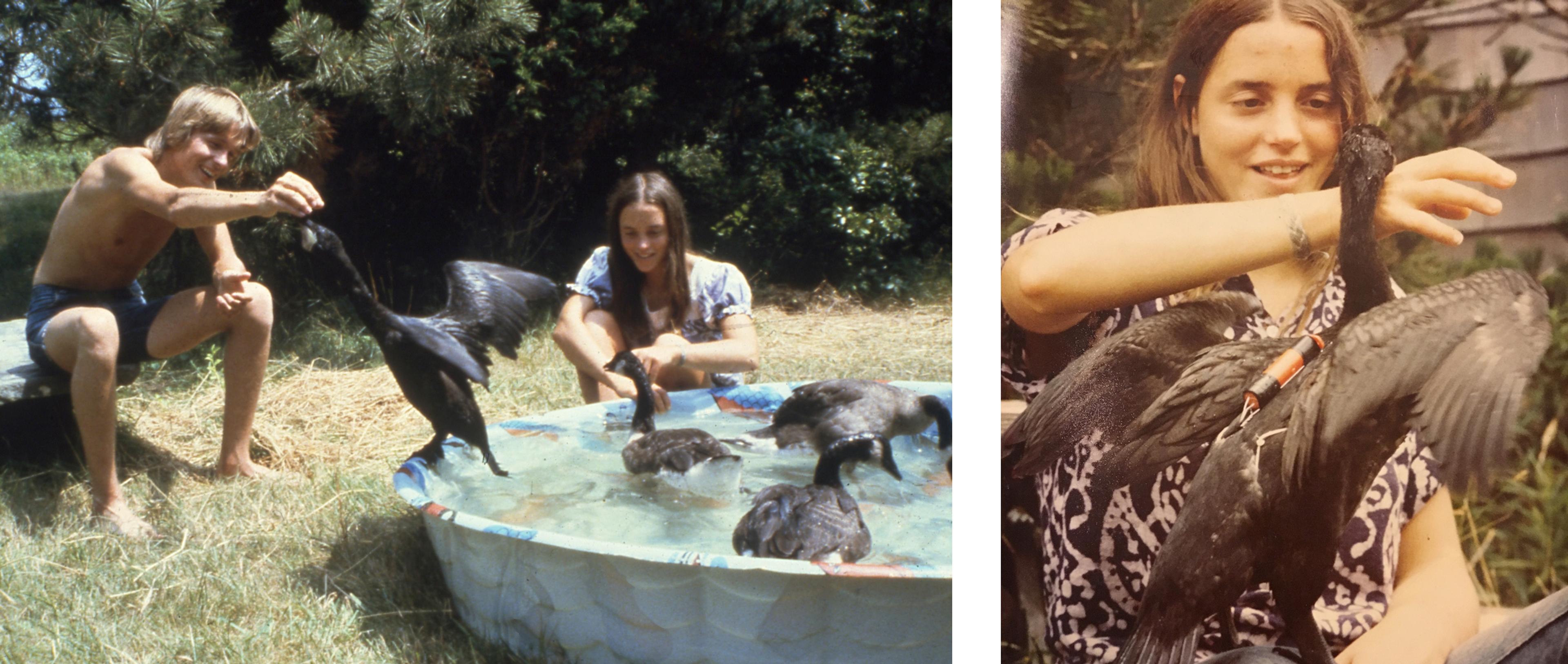
Left photo: Me and Geir Gabrielsen, then my dad's student, in our back yard in Woods Hole in the summer of 1979. We raised baby cormorants at home, and then used acoustic heart rate transmitters (right photo) made by my dad to measure heart rate in cormorants when they dove voluntarily. We showed that "diving bradycardia", previously thought to be an adaptation to diving, was largely a fear response that resulted when scientists forcibly submeged animals. When our home-reared cormorants swam freely with us they showed very little "diving bradycardia". Geir is now an exotoxicologist at the Norwegian Polar Institute.
Adventures in Norway
In fact, an important part of my introduction to science took place in Norway. Long after my dad had pissed off pretty much all of his colleagues in the U.S., he still had scientific friends in Norway, and he travelled there regularly to collaborate with them. I first visited Norway when he brought our family on one of these trips. He bought an old Norwegian fishing boat in ill repair named the "Nordlys." I remember boarding the boat in Bergen harbor, where we feasted on smoked mackerel from the fish market and then headed down the coast on a voyage that was glorious, memorable and quite dangerous.
A few years later my dad was planning an expedition to study ptarmigans on the island of Karlsoy near Tromso with Geir and several other scientists. I wanted to join, but there was no funding to bring an unskilled 17-year-old along, and flights to Norway were expensive. So, I got a cheap flight to Amsterdam, where I bought a bicycle and made my way to Tromso by a combination of pedal-power and train, including a thrilling bike ride over the Dovrefjell from Oslo to Trondheim. On Karlsoy, we lived in an old farmhouse, and went tromping across the island under the midnight sun to run field experiments on nesting ptarmigans.
Working in Molly Potters lab
As an undergrad at Massachusetts Institute of Technology (MIT) majoring in biology, I struggled. Despite my privileged early exposure to science, I had not learned much in public high school and I was simply not prepared for MIT. I worked in a lab studying differentiation of blood cells, but I did not enjoy killing a mouse for each experiment. So, I sought refuge in a department where they did not kill their subjects: the (then) MIT Psychology Department. There I worked in the lab of Molly Potter, a towering intellect of cognitive psychology and also a warm, fun, and supportive mentor who fished me off the bottom of the wait list for grad school a year later. My dad was scandalized that I planned to study psychology, which in his mind had all the rigor of astrology.
But my dad was wrong. I learned from Molly how to make powerful inferences about the inner workings of the mind from humble behavioral data, which is a bit like trying to figure out how a car works just by driving it around. Still, in cognitive science, as in auto mechanics, there is no substitute for looking under the hood. During my first year in grad school, the first noninvasive brain imaging study of human visual cortex was published on the cover of Science magazine, showing a very blurry yellow blob at the back of the head when people looked at patterned visual stimuli compared to diffuse illumination.
I was blown away. I wrote a proposal to use this device to answer a suite of questions about the mind, and sent it to all the brain imaging labs in the world (there were four). Does mental imagery engage the same brain machinery as visual perception? Does attention modulate responses early in the visual processing pathway? Where in the brain do we match incoming visual information with stored descriptions of what familiar objects look like? I gave a draft of the proposal to Molly, and she was furious. In her mind I was "selling out" to neuroscientists who failed to understand the power of behavioral data in revealing the mechanisms of the mind. But she got over herself the next day and has supported my efforts to answer cognitive questions with brain data ever since.
"I got frustrated and dropped out of graduate school three times to pursue journalism instead".
The luck turned
Only one of the brain imaging labs wrote back, and soon thereafter the trail went cold. Meanwhile I ran behavioral experiments on sentence understanding and visual perception. The questions were exciting and the experimental logic appealing, but most of my experiments bombed. For years. I got frustrated and dropped out of graduate school three times to pursue journalism instead. I once spent a month in Nicaragua at the peak of the contra war hitchhiking around in army jeeps and interviewing Sandinista officials in my abysmal Spanish. Molly was ever patient, insisting that my experimental ideas were good and that I was just unlucky with the data.
Eventually my luck turned, and a weird but powerful perceptual phenomenon landed in my lap. When people read strings of words presented rapidly in "rapid serial visual presentation," a method Molly had pioneered, they failed to see the second occurrence of a repeated word (even when several other words intervened). Molly and her colleague Helene Intraub, who had first discovered this phenomenon, kindly allowed me to try to get to the bottom of it for my Ph.D. thesis. Nine months and seventeen experiments later I handed in my thesis on "repetition blindness." To celebrate, I spent the next four months travelling in Nepal and Indonesia.
The fun and success of working on a newly-discovered and whopping perceptual effect arrived after I had already decided that I was not going to make it in science. Meanwhile, important world problems loomed, and I spent the next year studying nuclear strategy with a MacArthur Foundation Fellowship in Peace and International Security, trying to diagnose the cognitive biases that perpetuated misconceptions about U.S. foreign and military policy. Although fascinating, I eventually decided that cognitive biases were not the primary drivers of bad policy, and it made more sense to pursue political activism and science separately.
I applied for and received an NIH "FIRST" Award to continue my work on repetition blindness. The brilliant Anne Treisman kindly enabled me to bring this work to Berkeley where I pursued the connections between repetition blindness (the failure to link one perceptual feature with two distinct object representations) and Anne's work on illusory conjunctions (the failure to correctly link two features to a single object representation). That work went well enough that I was offered a faculty position at UCLA a couple years later. I think I had a grand total of two publications at the time. It was so much easier back then to land a faculty position! One of my future colleagues explained to me that they had tried but failed to hire the great Mike Posner, and I was their "poor man's Mike Posner." I was flattered.
My first brain imaging experiment
All this time I had been trying unsuccessfully to claw my way into brain imaging centers to ask questions about perception using brain data. Then in my first semester at UCLA I got a phone call from John Mazziotta, the scientist who had published the paper in Science that blew me away in grad school. John ran the UCLA PET imaging lab and said that they needed a psychologist to consult on a grant. I agreed and reminded him that the experiments I had pitched to him long ago had still not been done and still had great potential. This back and forth went on for the next year, until one day John called to say that he needed a letter of collaboration faxed over that afternoon. I said, John, give me two subjects! That was how I got to do my first brain imaging experiment, a decade after I first started angling for the opportunity.
"He said 'it’s me or the scanner'. I chose the scanner".
Right around that time a new method was gaining steam that appeared to have many benefits over PET imaging. Functional MRI (fMRI) was much cheaper and did not use ionizing radiation, which meant that you could scan the same subject as many times as you liked. The center of the action was back in Boston at the Martinos Center. I turned down near-certain tenure at UCLA to take near-certain non-tenure at Harvard to try to gain access to fMRI equipment. Even so, it was over a year before I heard back from the director of the Martinos Center, with an offer that I could use the scanner Saturday mornings from 6 to 9 am. My partner lived in D.C., and we saw each other only on weekends. He said 'it’s me or the scanner'. I chose the scanner. He buckled. Phew!
Grad students at Harvard knew that junior faculty were temp workers with lower job security than the janitors, so they worked mostly with the fancy senior professors. But I managed to convince one brilliant undergrad (Josh McDermott) and a fantastic postdoc from another lab (Marvin Chun) to work with me on the side. I would pick them up before dawn in Harvard Square and we would head over to the Martinos Center to run experiments. We had no idea what we were doing. But when we got stuck, the indefatigable Ken Kwong (who pretty much invented fMRI, and who seemed to be always present at the Martinos Center, no matter what hour) would come into the scanner control room and calmly announce, "I think you broke the scanner. Let me see if I can fix it!"
We eventually learned that when the message "arcing may occur" came across the scanner console, we were on the right track.

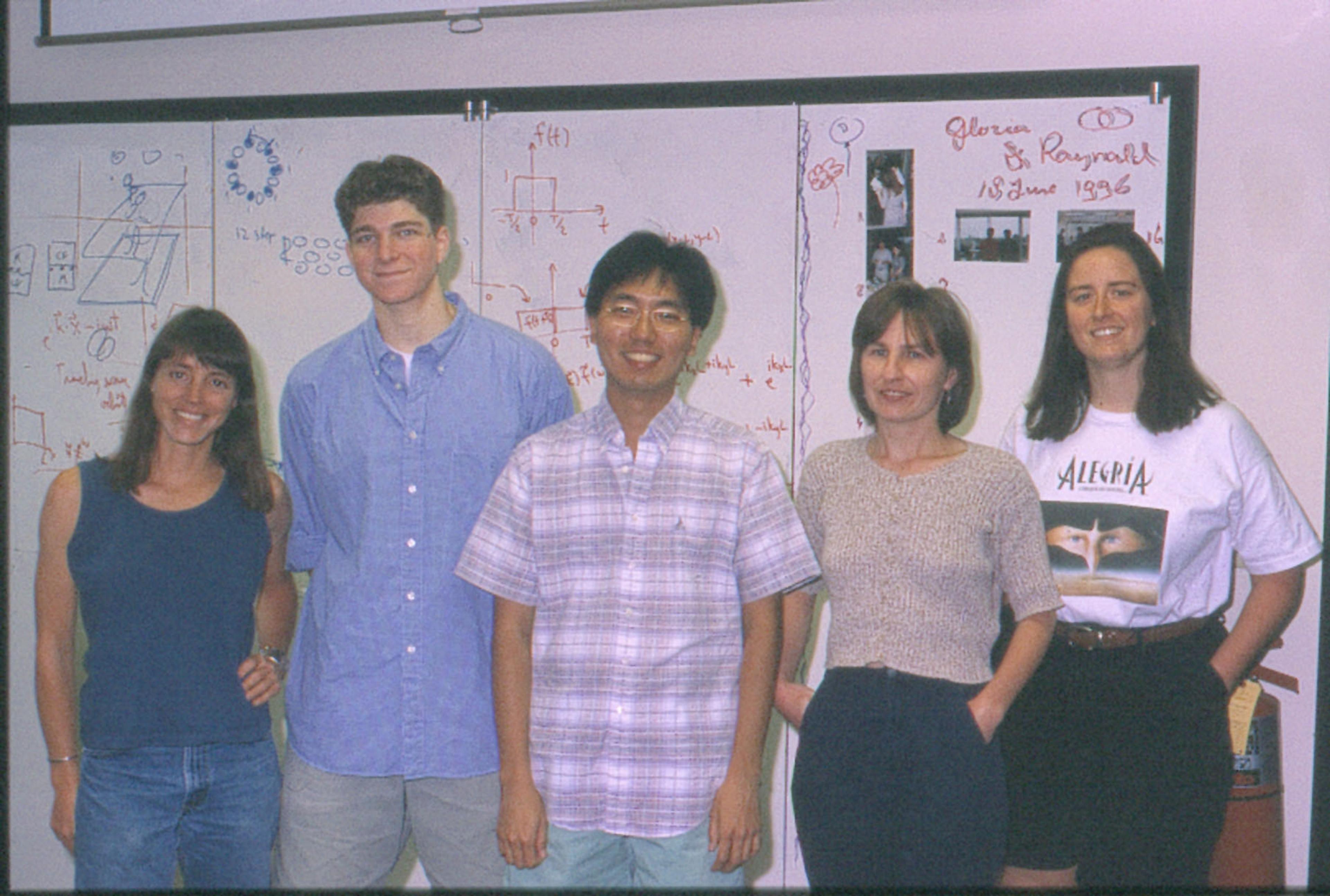
The Kanwisher lab circa 1996. From left to right: Nancy, Josh McDermott (then an undergrad), Marvin Chun (postdoc), Ewa Wojciulik (postdoc), and Jody Culham (grad student).
Problem solving
For a while we tried to study object perception, but our findings were not very robust. We needed a result, and fast, or we would lose our precious access to the scanner. If there was one functional response that almost had to be present in the brain, but had not been clearly described with brain imaging, it was a specialized circuit for face recognition. So, we scanned people while they looked at faces and while they looked at objects, and we asked if any brain regions responded more to the faces than objects. (Not rocket science!) Indeed, almost every subject showed at least one nice activation blob, where we could see in the raw time course of the fMRI response a big response during blocks of faces and a much lower respond during blocks of objects.
To publish this result, we needed to solve a few problems. First, what was most consistent across subjects in the pattern of face activations? The challenge is that different people's brains are quite different anatomically, so it is not clear what counts as the "same place" in two different brains.
"This enabled me to analyze the data in a way I understood, using ... Excel (!)"
So, we taped pictures of each person's activation pattern up on a long wall and spent days walking back and forth staring at the patterns, letting our own visual systems extract what was common across subjects. Indeed, each participant showed a face-selective response in an approximately similar location on the bottom of the right hemisphere just above the cerebellum. Our second problem was that these activation maps were computed using in-house software from the Martinos Center that did not account for the large number of statistical tests we were performing (on each of the tens of thousands of 3D pixels or "voxels" in our images). The standard methods for dealing with this problem ("Bonferroni correction") seemed too stringent, but the main alternative method devised by a scientist in the UK was impossible to understand. So, I decided to do something I did understand: use half the data within each subject to find the relevant voxels, and then measure their response in independent data. This enabled me to analyze the data in a way I understood, using ... Excel (!).
It also had the great advantage that we could systematically study regions with the same functional response profile across participants even if the region did not land in the same location defined by anatomy alone. This "functional region of interest" (fROI) method had been used earlier in studies of a brain region that processes visual motion by Roger Tootell and his colleagues. We developed it further and went to town.
Fruitful cooperation
Over the next few years, we used the fROI method to run dozens of control experiments, establishing that the fusiform face area (FFA) was detectable in nearly every normal subject, and truly did respond very selectively to faces. Galit Yovel and I showed that it was sensitive to individual identity in upright faces, but not inverted faces (echoing much earlier behavioral findings), Frank Tong and I showed that its activity was correlated with perceptual awareness of faces even when the stimulus did not change (in binocular rivalry), and Kathy O'Craven and I showed that you could turn on this region just by imagining faces with your eyes closed. More recently we and others have shown that electrical stimulation of this region produces a percept of a face, and Heather Kosakowski and Rebecca Saxe kindly allowed me to kibbitz with them in a study showing that this region it is present in 6-month-old infants. Artificial neural networks are also proving surprisingly informative: Ratan Murty and I showed that they can very accurately predict the FFA response to new stimuli, and Katharina Dobs showed that face-selective regions arise spontaneously in networks trained on both faces and objects, providing a strong hint about why the brain has an FFA in the first place.
"Imagine my luck to have these incredible scientists just down the hall!"
Just as importantly, the fROI method that we developed and promoted for the FFA proved a powerful way to discover and study in detail many other functionally distinctive regions of the cortex, including those that respond selectively to scenes, bodies, and text, as well as regions that respond during quintessentially human functions like perceiving music, understanding language and thinking about other people's thoughts. Each of those new directions took me to corners of the field I knew nothing about, and I could only do this work because I was lucky to have brilliant colleagues who were experts in these topics.
Josh McDermott, who had worked with me on the first paper on the FFA when he was an undergraduate (see photo above), and who is now a world expert on hearing, was central to the work on music. Rebecca Saxe taught herself about social cognition as a grad student, enabling her to discover a cortical region that turns on only when you consider what someone else is thinking. And Evelina Fedorenko applied the fROI method to answer a centuries-old question by showing that language and thought are distinct in the brain. Imagine my luck to have these incredible scientists just down the hall! I still have to pinch myself to believe it.


A four-generation scientific matriarchy. From right to left: My brilliant mentor and Ph.D. advisor Molly Potter, me, my superstar former Ph.D. student (and now colleague) Rebecca Saxe, on the day of the Ph.D. thesis defense of her own brilliant student Liane Young (now a professor at Boston College).
Revealing the architecture of the human mind
To me the most exciting aspect of this work is that it reveals the basic architecture of the human mind. Perhaps it is a good thing I did not gain access to brain imaging equipment when I first sought it. In the intervening decade I learned more about cognitive science, and developed a deeper appreciation of the theoretical debates and unsolved mysteries of the human mind. The gift from this period was a lifetime of questions about the mind that I could tackle by measuring activity in the brain.
But exhilarating as this adventure has been, the methods available in humans suffer from serious limitations. We cannot see the neural code, discover the connectivity, or causally intervene on the brain in humans with anything like the precision that is possible in animal studies. So, it was a special thrill for me when Doris and Winrich found face patches in macaques using fMRI, and went on to nail each of these fundamental questions in monkeys that I could not answer in humans. They are scientific heroes to me, and it is a thrill to receive the Kavli Prize in neuroscience jointly with them.